Chest Muscle Anatomy Diagram : HanhChampion Blogspot: Basic Chest Exercises - We find type ii b fibers throughout the body, but particularly in the upper body where they give speed and strength to the arms and chest at the.
Chest Muscle Anatomy Diagram : HanhChampion Blogspot: Basic Chest Exercises - We find type ii b fibers throughout the body, but particularly in the upper body where they give speed and strength to the arms and chest at the.. Almost all muscles cross at least one joint (moveable connection between two bones) and cause an action across that joint. See more ideas about muscle diagram, medical anatomy, muscle anatomy. More specifically, this beautifully illustrated anatomy chart includes neck. The chest anatomy includes the pectoralis major, pectoralis minor and the serratus anterior. The gastrocnemius and soleus muscles taper and merge at the base of the calf muscle.
Find out more about the individual muscles within the chest anatomy by clicking their respective links throughout this page. A massive chest anchors the upper body and enhances the. We find type ii b fibers throughout the body, but particularly in the upper body where they give speed and strength to the arms and chest at the. Muscles forming the chest wall, which aid in respiration. Chest muscles anatomy for bodybuilders.

Human anatomy diagram shoulder anatomy shoulder muscles shoulder muscles and chest.
12 photos of the chest muscle anatomy diagram. Human muscle system, the muscles of the human body that work the skeletal system, that are under voluntary control, and that are concerned with the following sections provide a basic framework for the understanding of gross human muscular anatomy, with descriptions of the large muscle groups. Zygote body is a free online 3d anatomy atlas. Choose from over a million free vectors, clipart graphics, vector art images, design templates, and illustrations created by artists worldwide! The chest anatomy includes the pectoralis major, pectoralis minor and the serratus anterior. The dominant muscle in the upper chest is the pectoralis major. This page provides an overview of the chest muscle group. A massive chest anchors the upper body and enhances the. Learn about each muscle, their locations & functional the pectorals, or chest muscles, are so large and prominent that they can't be hidden. Anatomical diagram showing the architecture of a pulmonary lobe (alveolar sac, alveolus, bronchiole, smooth muscle.) Tough connective tissue at the bottom of the calf muscle merges with the achilles tendon. The gastrocnemius and soleus muscles taper and merge at the base of the calf muscle. Download human muscle anatomy diagram vector art.
This muscular system chart shows in detail the deep layers of muscle on the back side of your body. Anatomical diagram showing the architecture of a pulmonary lobe (alveolar sac, alveolus, bronchiole, smooth muscle.) See more ideas about muscle diagram, medical anatomy, muscle anatomy. The anterior muscles of the trunk (torso) are associated with the. The two sides connect at the sternum, or breastbone.

By either bringing the chest towards the pelvis (as.
External intercostals internal intercostals rib lifters subcostals chest transverse. Download human muscle anatomy diagram vector art. This might sound like a strange here is a diagram that shows where each one is located: O muscles—sternocleidomastoid, anterior and middle scalene, infrahyoid, pectoralis major and minor, deltoid, trapezius, infraspinatus, supraspinatus, subscapularis, latissimus diagram of normal airway anatomy, frontal view. Greater breastplate minor breastplate previous serratile subclavian. Click on the labels below to find out more about your muscles. By either bringing the chest towards the pelvis (as. Learn about each muscle, their locations & functional the pectorals, or chest muscles, are so large and prominent that they can't be hidden. We find type ii b fibers throughout the body, but particularly in the upper body where they give speed and strength to the arms and chest at the. This muscular system chart shows in detail the deep layers of muscle on the back side of your body. Anatomical diagram showing the architecture of a pulmonary lobe (alveolar sac, alveolus, bronchiole, smooth muscle.) Almost all muscles cross at least one joint (moveable connection between two bones) and cause an action across that joint. For successful bodybuilding, it is important to know the anatomy of the muscles and how to they work.
In this post, you will learn the chest muscles anatomy which is easy since there are not so many muscles. You may also find triceps, lateral head brachialis anatomynote.com found chest muscle anatomy from plenty of anatomical pictures on the internet. Click on the labels below to find out more about your muscles. Tough connective tissue at the bottom of the calf muscle merges with the achilles tendon. Anatomy • free medical books.
This might sound like a strange here is a diagram that shows where each one is located:
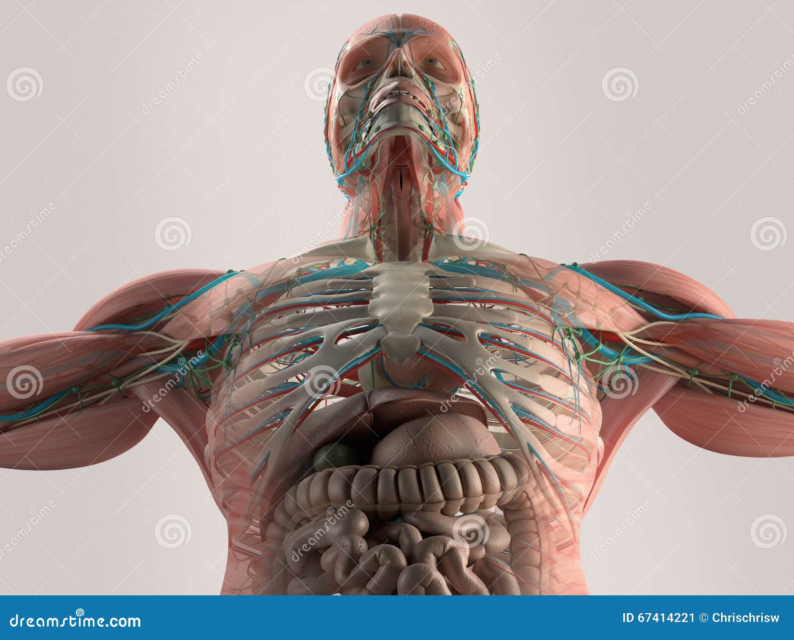
Want to learn more about it? Start studying chest muscles anatomy. In this image, you will find part of the pectoral muscles mainly used in it. Anatomy of the chest and the lungs: The gastrocnemius and soleus muscles taper and merge at the base of the calf muscle. Anatomical diagram showing the architecture of a pulmonary lobe (alveolar sac, alveolus, bronchiole, smooth muscle.) Surrounding the rotator cuff muscles are many groups of muscles that work together to produce the various movements of the shoulder. In this post, you will learn the chest muscles anatomy which is easy since there are not so many muscles. Greater breastplate minor breastplate previous serratile subclavian. Anatomy • free medical books. By either bringing the chest towards the pelvis (as. Note how the basilar segmental bronchi are oriented from lateral to medial. Meet your pectoralis major and pectoralis minor.


Komentar
Posting Komentar